
Artificial intelligence predicts breast cancer…a new technology that outperforms traditional methods

Artificial intelligence predicts breast cancer…a new technology that outperforms traditional methods
With the escalation of health challenges globally – from the increase in cases ofbreast cancer to the spread of chronic diseases – there is an urgent need to adopt more accurate diagnostic tools capable of detecting danger in its early stages before symptoms worsen. At the heart of this scene is the third goal of the Sustainable Development Goals (SDGs) (good health and well-being), which emphasizes the importance of prevention, improving the quality of examinations, facilitating access to health care services, and enabling individuals to understand their health risks more clearly.
The problem of late diagnosis of breast cancer is one of the most prominent health challenges in our societies, as delayed detection of the disease leads to weak treatment effectiveness and complicates recovery paths. In this context, artificial intelligence is advancing as one of the most important emerging medical tools, with its ability to analyze images and data with accuracy that exceeds traditional human capabilities.
Preliminary results issued by a research team at Harvard University on the Clairity Breast model provide a remarkable addition in this field. The model reveals an outstanding ability to predict the risk of breast cancer before any symptoms appear, clearly superior to traditional indicators used in risk assessment.
Excellence in detecting breast cancer
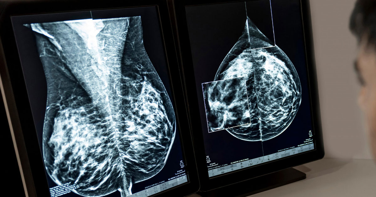
The American Cancer Society recommends that women at average risk begin annual mammograms (a specific type of X-ray designed to image breast tissue with high resolution) starting at age 40. However, recent studies indicate that women under the age of 40 constitute the fastest-growing group in breast cancer diagnosis rates, including advanced cases, which highlights the need to improve the accuracy of current assessments and reconsider the timing of traditional examinations.
In the context of seeking to bridge the early detection gap and improve diagnostic accuracy, the preliminary results of this research team reveal an advanced artificial intelligence model – based on the analysis of mammogram images only – capable of predicting the risk of breast cancer during the next five years with an accuracy far superior to traditional methods, especially those that rely on breast tissue density alone.

This model is distinguished by its ability to capture precise patterns in breast tissue that radiologists cannot see with the naked eye, which allows for a more accurate and clearer classification of risk levels among women.
Dr. Constance Lehman, a professor of radiology at Harvard University and the study’s lead researcher, explains that existing risk assessment methods – such as age, family history, genetic factors, and breast density – are no longer sufficient for reliable early detection.
Advanced capabilities for early detection
The Clairity Breast model is the first FDA-approved AI model based entirely on image analysis. It was trained on more than 421,000 mammogram images collected from 27 medical facilities in Europe, South America, and the United States.
Training was based on images of women who later developed breast cancer and those who did not over the next five years, enabling the model to recognize subtle patterns and subtle changes in breast tissue associated with a higher risk. It was also calibrated on an independent test set using a deep neural network capable of producing accurate predictions of infection probabilities over five years.

The research team explained that the artificial intelligence model has the ability to monitor subtle changes in breast tissue that cannot be observed by the human eye, which makes it a qualitative leap in the level of medical analysis. Because this model opens new horizons for using hidden information within radiological images – that could not be exploited by traditional methods – and it is not limited to improving the accuracy of diagnosis, which reflects the great development that artificial intelligence has begun to provide in the medical field and improving the efficiency of health services and making them more sustainable.
More accurate analysis of breast tissue reveals true risk disparity
The artificial intelligence model was put to the test with a vast collection of more than 236,000 bilateral mammogram images from five medical centers in the United States, in addition to 8,810 images from one European center. Radiologist-determined breast density data (dense vs. non-dense breasts), along with 5-year breast cancer outcomes, were collected from medical and tumor registries.
In this, the study team relied on classifying breast risks – according to the National Comprehensive Cancer Network – into three categories: low, medium, and high. When the results were analyzed using the artificial intelligence tool, it was found that women in the “high risk” category were four times more likely to develop breast cancer compared to those in the “low risk” category. On the other hand, traditional detection tools showed a very small difference between the two categories, not exceeding 0.5%, which highlights the accuracy of artificial intelligence in estimating danger.

Concerning this, Christiane Cole, Director of the Department of Diagnostic and Interventional Radiology at Aachen University Hospital (RWTH), located in western Germany, said: “The results of this large-scale analysis show that artificial intelligence models serve a more accurate and stronger classification of breast cancer risk than relying on breast density alone.”
The future of breast cancer risk assessment
We must be aware that breast density legislation – implemented in 32 US states – requires health care providers to inform women of breast density results when undergoing a mammogram, in an attempt to raise awareness of the factors affecting the risk of breast cancer. Although this step is important, it is not sufficient. Breast density is no longer an accurate indicator of risk.
So AI introduced this technology based on risk assessment based on analysis of mammogram images, which is a real shift in early screening methodologies; Thus, women who are most at risk of developing breast cancer can be identified with accuracy beyond traditional methods. This allows examinations to begin at an early age when necessary, instead of strictly adhering to the currently approved age.
The study team also emphasizes that the future of screening must integrate risk assessment based on artificial intelligence. Because it provides deeper information, and the examination is not limited to assessing breast density only, to help women understand their health risks in a clearer and more realistic way.
Such developments represent an important step towards enhancing prevention, and one of the essential pillars of the third goal of the Sustainable Development Goals (SDGs) related to good health and well-being, by enabling women to obtain early diagnosis, and supporting more fair, accurate and proactive health systems.
Also, this progress in using artificial intelligence for early detection of breast cancer reveals a fundamental shift in the health sector, from late diagnosis to proactive prevention, which is directly in line with the Sustainable Development Goals (SDGs), especially Goal No. (3) Good Health and Well-Being, which seeks to reduce the burden on health systems.
Therefore,The Earth Guards Foundationsees that promoting these technologies represents a pivotal step in building more equitable and efficient health systems, especially in countries that suffer from limited resources and delayed diagnosis. Therefore, the Foundation calls for supporting the adoption of these smart tools, training medical personnel on them, and expanding early breast cancer screening programs, as a direct investment in women’s health, and in a more sustainable future capable of protecting lives.


