

One image may reveal the chances of survival.. How does artificial intelligence help in choosing cancer treatment?

One image may reveal the chances of survival.. How does artificial intelligence help in choosing cancer treatment?
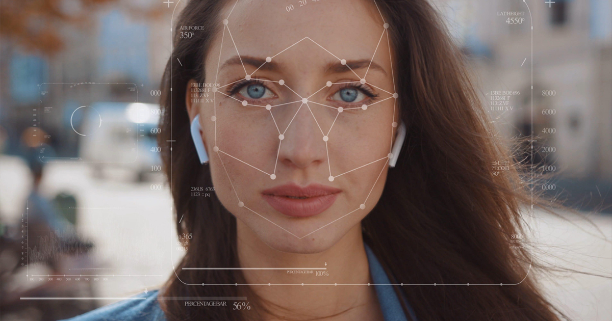
What if a doctor could learn more about a cancer patient's ability to tolerate treatment from a regular photo of his or her face? The idea may seem akin to science fiction, but it became the focus of a recent medical study, during which researchers developed a system based on… artificial intelligence To analyze facial features and extract indicators related to biological age and death risks in patients.
The innovation came from an institution Mass General Brigham American Medical Center, and its results were published in the journal "The Lancet Digital Health", opening a new field for developing tools that may help doctors in the future make treatment decisions that are more appropriate to the physical condition of each patient.
The importance of the study lies in a question that goes beyond the capabilities of technology: Can information extracted from a simple image contribute to improving the quality of health care, especially when the doctor is faced with a decision related to intensive treatment or major surgery?
How does artificial intelligence read facial features?
The researchers developed a baseline model that was trained on more than 40 million images of faces, spanning diverse age groups and ethnic backgrounds, with the goal of learning visual patterns that could be used to assess health-related indicators.
From this model, the team derived two complementary tools; The first"FAHR-FaceAge", which estimates the patient's apparent biological age from his facial features, and the second "FAHR-FaceSurvival", Which estimates the risk of death directly from the image, without first calculating biological age.

Although the two tools start from the same model, each captures different information. The first focuses on the folds extending from the nose to both sides of the mouth, and the forehead area above the eye sockets and temples, while the second analyzes the areas below the eyes and around the bridge of the nose.
Researcher Hugo Aerts, one of the senior supervisors of the study, explained that training a basic model of this size allows the development of multiple health indicators from images of faces, rather than creating an independent model from scratch for each new medical use.
What did the results reveal?
Researchers tested the relationship between apparent age estimates and health outcomes in cancer patients who received radiation therapy in the United States, over an average follow-up period of four years.
The results showed that patients whose apparent age was estimated by the tool to be five or more years older than their actual age had a 21% higher risk of death compared to the reference group.
In contrast, the relative risk was reduced by 17% in patients who looked five or more years younger than their actual age.
These percentages express statistical differences between the study groups, and do not mean that the system can determine the date of death of a particular person or guarantee his survival.

The researchers also monitored the association of smoking, excessive alcohol consumption, and drug use with indicators of apparent aging, while the age estimation tool maintained relatively stable results despite differences in lighting, photography angles, facial expressions, and makeup.
Estimates of apparent age were not significantly affected by most of the plastic surgeries the researchers tested, while slight differences emerged after rhinoplasty.
The results indicate that facial features may contain information related to general health status, which in the future will allow patients to be evaluated from a broader perspective than their ages recorded in medical files.
Does a facial image help in choosing treatment?
Before recommending intensive radiation therapy or major surgery, the doctor needs to estimate the patient's body's ability to tolerate the treatment and its possible complications.
Dr. Raymond Mack, a radiation oncologist at Mass General Brigham, believes that an aging population increases the need for more accurate methods of assessing physical reserve and physical weakness before making treatment decisions.
Hence the potential benefit of facial analysis; A normal photograph may provide an additional indicator to help evaluate health status, in addition to the usual medical examinations and assessments.

If a low-cost, easy-to-use tool could be developed for this purpose, would it help improve access to more accurate health assessments in the future?
The question opens up a development dimension related to the quality and accessibility of care, especially since medical innovation gains greater value when its benefits become accessible to diverse categories of patients.
This goal is consistent with Sustainable Development Goal 3, good health and well-being, especially Objective 3.8 associated with universal health coverage and the availability of good basic health services, but reaching this stage will require additional evidence confirming the accuracy of the technology and its usefulness in different medical settings.
When a facial image becomes medical information
The use of facial images in health risk assessment raises questions regarding patient privacy and the limits of relying on results produced by intelligent systems.
The image that may help the doctor understand the physical condition also contains personal information that requires protection, and statistical estimates require medical interpretation that takes into account the patient’s circumstances and health history.

These considerations are particularly important because researchers still call for additional studies before the two tools can be used on a broad clinical scale.
Raymond Mack stressed that the goal of the technology is to support personal treatment decisions, while preserving the role of the doctor in interpreting the results and choosing the appropriate treatment.
This study reflects a growing trend in the use of artificial intelligence in medical image analysis. In another application, I addressed… The Earth Guards Foundation A technique based on x-ray images to predict the risk of breast cancer, demonstrating the diversity of indicators that smart tools can extract to support health risk assessment.
The future of care begins with understanding people
The new innovation provides insight into how ordinary images can be used to develop personalized medicine aids, but it puts researchers on the responsibility of proving its accuracy and effectiveness before introducing it into standard treatment practice.
The experience reveals a broader issue that it addresses The Earth Guards Foundation In its treatment of innovation and development: How can technological progress be transformed into a tangible health benefit, while preserving human privacy and his role in decisions that affect his life?
Artificial intelligence may be able to extract indicators that are difficult to notice with the naked eye, but the medical benefit from it will remain linked to scientific research, careful evaluation, and clinical judgment.
In the journey of treating a cancer patient, a facial image may one day become a source of additional information that helps the doctor, while the patient’s future remains too big to be summarized by a single image or algorithm result..


